

The incredible floating atom, pop art placentas and psychedelic bubbles: EPSRC science photography awards
The top prize went to a remarkable image of a single, floating atom captured by an Oxford University student using an ordinary DSLR camera.
Science and art are often seen as antithetical fields, however, photography can act as an effective bridge between the two disciplines, revealing the inherent beauty in many scientific pursuits.
In light of this, the Engineering and Physical Sciences Research Council (EPSRC) has announced the winners of its national science photography, with the top prize going to a remarkable image (see above) of a single, floating atom captured by an Oxford University student using an ordinary DSLR camera.
The stunning set of images features a variety of different scientific disciplines, ranging from biology and chemistry to robotics and the environmental sciences.
"The winners we have selected demonstrate how science and technology is affecting people's lives today at many different physical scales," said Ellie Cosgrave, a BBC broadcaster and judge on the competition. "We have examples from the nano-scale, how our cells are working; how our immune systems might be able to treat different types of diseases; all the way up to technologies that can help us move through cities."
The 100-plus photos were submitted by scientists conducting ground-breaking research being supported by the EPSRC.
"Not only do we have really strong, attractive photographs, the stories behind them about the research and why it is being done are inspiring," said Dame Ann Dowling, another judge. "Much of this work will lead to innovations that transform lives and, in this Year of Engineering, it's marvellous to see these great examples of transformational research."
To take his winning photo, David Nadlinger, a quantum physics PhD candidate, suspended a single strontium atom in the air between two metal electrodes – just two millimetres apart - and illuminated it with lasers. The atom, which is held in place by a strong magnetic field, is just about visible to the camera as a tiny purple-blue dot.
In atomic terms, strontium atoms are fairly large -measuring around 215 billionths of a millimetre across - but this is still infinitesimally small. So, how is it possible that the atom is viewable with the naked eye in this image?
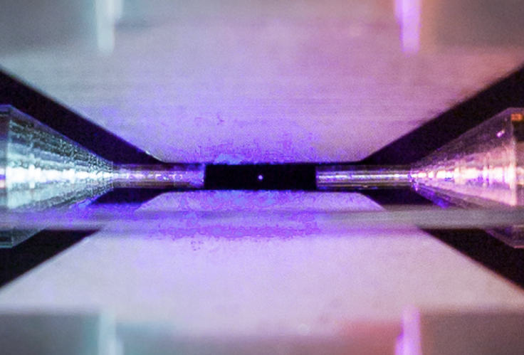
"When illuminated by a laser of the right blue-violet colour, the atom absorbs and re-emits light particles sufficiently quickly for an ordinary camera to capture it in a long exposure photograph," according to the EPSRC.
In other words, we are not seeing the true outline of the atom, but rather the laser light that it is reflecting.
Explaining how he came up with the idea for the photo Nadlinger said: "The idea of being able to see a single atom with the naked eye had struck me as a wonderfully direct and visceral bridge between the miniscule quantum world and our macroscopic reality."
"A back-of-the-envelope calculation showed the numbers to be on my side, and when I set off to the lab with camera and tripods one quiet Sunday afternoon, I was rewarded with this particular picture of a small, pale blue dot."
In terms of camera equipment, Nadlinger's setup was fairly basic apart from his use of extension tubes – an accessory which increases the focal length of a lens that is useful for extreme close-up photography.
See the best of the rest of the images below:
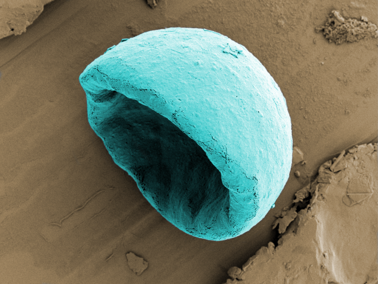
Biodegradable, bowl-shaped micro-particles, such as these, could help to fight stubborn cancers. Unlike healthy organs, tumours do not have an extensive network of blood vessels making them difficult to treat due to the fact that anti-tumour drugs cannot penetrate very far. However, these minuscule 'micro-bowls' can be injected alongside cancer drugs to help them penetrate further into the tumour. After being injected, the patient receives a dose of ultrasound which causes tiny micro-bubbles, which become trapped in the cavity of the bowl, to vibrate. The surrounding fluid is rapidly moved around by the vibrations, forcing the bowl into the tumour, while taking the drug along with it. The image was taken using a Zeiss Merlin Compact Field Emission Gun at the Dunn School of Pathology and was colourised using Adobe Photoshop.
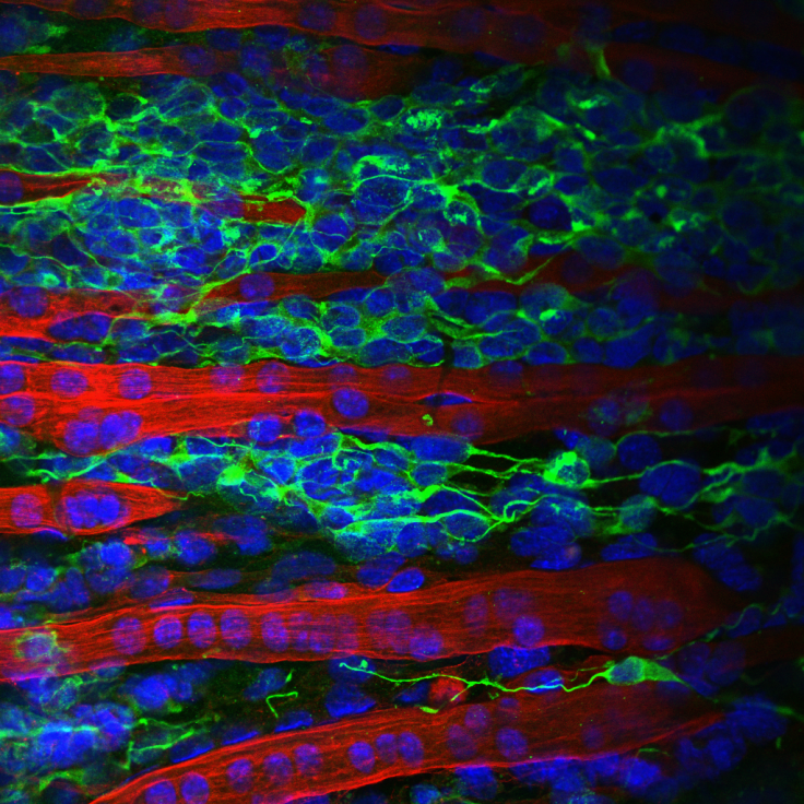
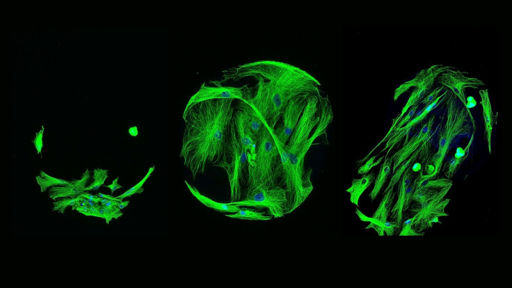
In order to understand how neurodegenerative disorders, such as Parkinson's or Alzheimer's progress, researchers need to generate biologically accurate models of the regions in the body where the nerves and muscles interact, to enable new treatments to be tested. Traditionally this testing is done using animal studies which are often ethically questionable and do not always accurately represent the unique responses of the human body. This research is focused on developing biological models of the nerve-muscle interface which could, one day, remove the need for animal testing in this field. The image shows a cross-section of the connections between artificially engineered nerve cells (neurons) and muscle fibres. The elongated red structures are muscle cells, whilst the green structures are neurons. Taken at Queens Medical Centre, Nottingham using a Zeiss LSM880 confocal microscope.
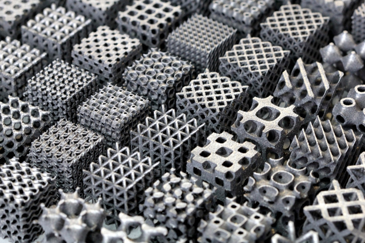
These impressive aluminium lattice structures are manufactured using a specialised type of 3D-printing known as selective laser melting. Their unique shapes lend them incredible strength and stiffness, enabling engineers to significantly minimise the weight of components without compromising on strength or performance. This is important for many sectors, such as the aerospace and automotive industries, because weight directly correlates to the fuel efficiency and environmental impact of travel. Taken using a Canon 60D DSLR, with the exposure tweaked using post-processing software.
Determining the ability of cells to attach to materials is an essential step towards the development of bio-materials that will allow researchers to effectively grow stem cells. Stem cells can be turned into any type of cell in the body, meaning they hold potential for treating a host of debilitating diseases. This imaging method shown in this photo - known as high throughput screening - is being used, in this case, to screen hundreds of materials in order investigate their ability to influence the growth of stem cells into bone cells.
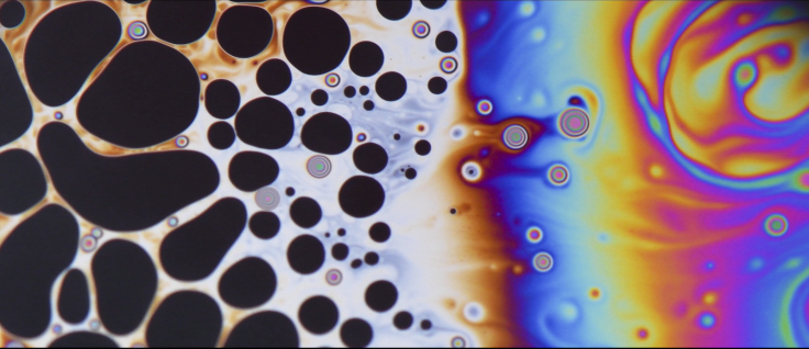
This wonderfully psychedelic image may look like it was generated with the help of mind-altering substances, however, the reality is rather more mundane. In actual fact, it's a close-up photo of fluid patterns on top of a spherical soap bubble in a kitchen sink. Taken using a Nikon D500 with a 105mm VR macro lens. The RAW image was then processed in Adobe Lightroom for exposure and level adjustments.
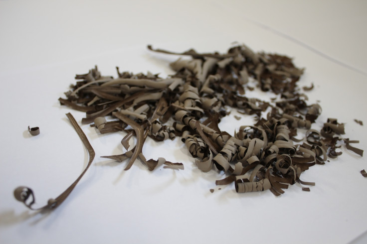
Due to its ubiquity, soil has the potential to become a useful construction material which could help house the world's population in a sustainable manner through the replacement of concrete blocks. However, to unlock its full potential, researchers are attempting to use various techniques to transform different soil clays into water-resistant, durable and strong materials. These clay ribbons, as an example, were refined from naturally occurring Sudanese soil sedimentation.

A farmer holds two sets of Lady's Fingers (Okra) in each hand, which were both grown for the same length of time. The smaller pods on the right were grown using manually irrigated crops, while the larger ones on the left were produced using an automated crop irrigation system which makes use of highly localised weather forecasts. This prototype system increased crop yields by 100% when tested, in addition to minimising labour needs and composting requirements. It also reduced water and energy consumption by 82%. This led to more efficient power use on the farm and higher revenues.
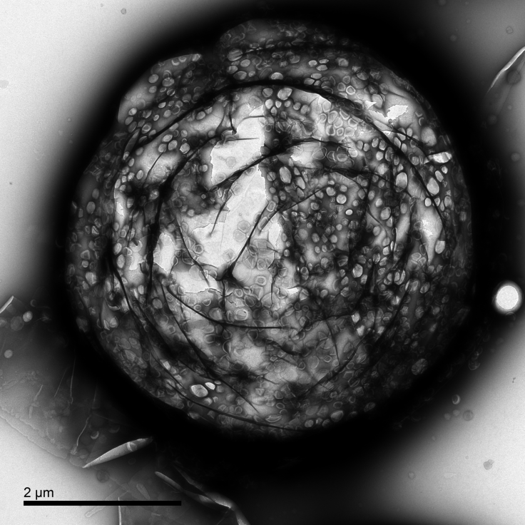
This image, taken with an electron microscope, shows a micro-bubble measuring just over four micrometres - one millionth of a metre - across. As mentioned above, these micro-bubbles are being explored for their use in improving the delivery of drugs which target tumours, but they are also being harnessed to improve the quality of ultrasound medical imaging - like the type used by midwives to view babies inside the womb.
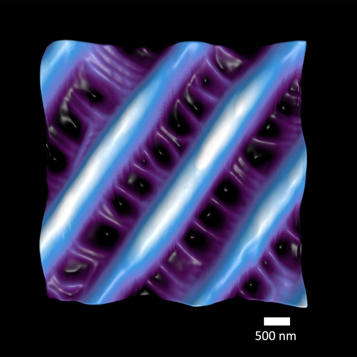
An extreme close-up of the surface of a butterfly's wing at the nanometre (one thousand-millionth of a metre) scale reveals its kaleidoscopic beauty. The image highlights tiny structures which trap the Sun's rays, producing a dazzling array of colours. It was taken using a Nanosurf Atomic Force Microscope which scans across a surface, detecting changes in height. It was then processed using Blender.

Placentas serve one important purpose - to support new life - however, they are fantastically diverse in shape and appearance. These images taken using high resolution photography show blood vessels which surround the growing foetus, which were then coloured in a Pop art style.
-

Football Clubs With The Most Arrests And Bans in 2022-23
-

Oil Prices Slip As Investors Eye Israel's Response To Iran Strike
-

Billionaire Declared Missing And Dead In 2018 Is 'Possibly Living In Moscow With Mistress'
-

Wendy's Sued For $20M After 11-Year-Old Gets Brain and Kidney Damage From 'Dirty Meal'


